
Accelerating and Democratizing Medical Innovation with Stratasys 3D Printing
Guest Blog: By Jack Stubbs, Associate Director, Human Performance in Healthcare, University of Central Florida
I started employing 3D printed prototyping over 20 years ago, in the mid-1990s, to introduce effective surgical device designs into a rapid development cycle.
We were able to test laparoscopic prototype devices with surgeons and OR personnel in simulated and animal test settings. Using Stratasys Dimension 760 and Dimension Elite FDM-based 3D printers, we could design and produce surgical devices literally overnight and have them ready for another round of testing. Conventional prototype approaches typically took weeks to produce. This work was accomplished within a small development company. Prior to purchasing the 3D printers, we had to send out all work to local fabrication shops, which was time-consuming and risky in terms of protecting our intellectual property. Bringing this capability in-house, the 3D printers paid for themselves in less than two months and enabled us to develop prototype fabrication as an additional business line.
3D printing capabilities have expanded significantly since then. Medical applications in particular have grown to include personalized prosthetic devices, casts for broken limbs, and implants for jaws, hips, and other skeletal structures. In the current decade, we have been using Stratasys’ Fortus 250mc and Objet260 Connex 3D Printers to produce 3D printed anatomical models and surgical training systems. With the new PolyJet materials that incorporate flexible and rigid within one build platform, simultaneously, we can design accurate anatomical and surgical skill trainers. These models could take weeks or months to produce with conventional subtractive approaches, but they are quickly fabricated with 3D printing.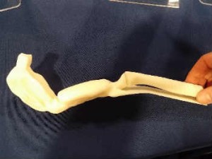
Recently, we have been working with Stratasys and Vital Images to develop approaches to produce anatomically correct 3D printed models from MRI and CT scans. Using Vitrea software, we can segment tissues from the DICOM stack (the file format for medical imaging) and directly export STL files (the file format for 3D printing) to 3D print anatomical parts. Advances in software and automation tools have significantly accelerated this process. For an Airway Intubation training model, we were able to print a full-scale example of the negative airspace of a person within eight hours after the MRI scan.
The U.S. Army tasked our team at the University of Minnesota to improve on the mannequins currently used by developing an Airway Intubation training model. Current models approximate human anatomy but without full realism, resulting in a gap in the skills needed to intubate a patient in the field. Basing our model on MRI scans of human anatomy, and producing it with Stratasys’ multi-material 3D printing capability, we better replicated the complexities of the real human.
3D printing also provides the flexibility to customize the model without increasing the per model costs. For example, you can incorporate sensors as needed to measure trainee performance and provide real time feedback, or create variations between models to test the full range of anatomy that one would encounter clinically. In the airway model, we created different versions of the model to simulate burn victims and patients with unusual mouth and face anatomy.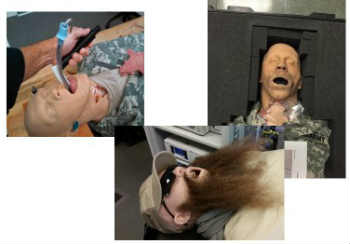
From left to right: basic intubation trainer, trainer with small mouth and chin, trainer with burn injuries.
Six of these airway training models have been created and provided to the U.S. Army for training. Initial feedback is positive. Using anatomically accurate 3D printed models enables trainees to develop the skills they need to intubate patients in the field without relying on training on a friend, animal or in the ER/OR on a patient who needs quick medical care. It allows trainees to practice over and over again to refine their medical technique. The 3D printed models are closer to actual human anatomy than any other trainer available and provide a positive training scenario that can be repeated, corrected and improved with feedback and guidance.
Within universities, there are limited facilities for fabrication. Owning a 3D printer provides the ability to fabricate models very quickly. We don’t need the large space and dedicated machinist typically required to operate conventional equipment. This also gives students the opportunity to design and experience rapid modeling and fabrication.
Finally, an exciting aspect of 3D printing is how it makes innovation and product development so accessible. It allows people who don’t have a lot of experience or training in mechanical design to try new things. Rather than being restricted to “subtractive” thinking – how do you build something by removing material – you can focus on how the device should look and function.
There is a student in our lab, a linguistics major, who designed a new prototype for an otoscope teaching device. This device gives the user the ability to train and learn how to use the device – integrated sensors measure the position, angle and movement of the device during use. The student was able to use open source CAD software to quickly design the entire device with fasteners, electronics, plugs and optics to produce a working prototype design and then 3D print it on the Stratasys Fortus 250mc for use and demonstration.
Student-designed otoscope.
So 3D printing is empowering people who may never have been an inventor to create something new. That is the power of 3D printing for healthcare and other industries!
Watch Jack Stubbs’ keynote presentation at HIMSS16.
To keep up to date about the latest advances in 3D printing for medical applications, please click here to sign up for the Stratasys Medical Innovation Series.
Follow Stratasys Medical on our new LinkedIn Showcase page.
